

Kate Criswell, Postdoctoral Research Associate, writes:
One of the key features that distinguishes vertebrate animals from our invertebrate cousins (such as insects and molluscs) is a backbone, or a series of vertebrae that run the length of the body. These vertebrae can range in number from only nine in frogs to over 300 in elongate animals like snakes and eels! They are important for providing structural support for the head and limbs and protecting the spinal cord. I study how vertebrae develop in a cartilaginous fish, the little skate, and compare skate development to other vertebrates to understand how the backbone evolved.
It is relatively easy to study development in skates, compared to other cartilaginous fishes, because they are oviparous, or egg-laying. Skate egg cases are often called mermaid’s purses and are similar to chicken eggs in that one egg contains a single embryo connected to a yolk. Little skate embryos take 5-6 months to develop and then will hatch out fully formed, looking just like an adult. To study vertebral development in skates, we can cut a small window in the egg case and look at the embryo under a microscope.


Vertebrae form from repeated blocks of embryonic mesoderm called somites. As the embryo develops, cells from the bottom portions of the somites migrate to the middle of the embryo and condense around the spinal cord to form cartilage or bone. By using a tiny needle to inject dye into the somites, I can track these cells and determine where they end up in the eventual vertebrae.

In a recent study, I used two different colours of dye to track cells from neighbouring somites. I found that one somite doesn’t give rise to just one vertebra, but that the somites actually recombine as they migrate.
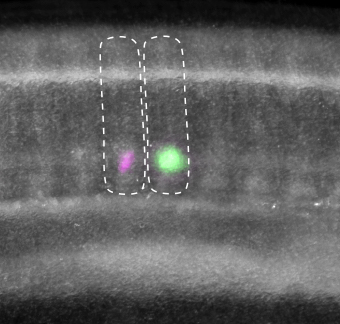

The result is that one vertebra contains cells from half of one somite and cells from half of the neighbouring somite. So, the backbone has segmented at two different points in its development: first, when the somites form, and second, during the migration process where the somite cells go on to form vertebrae. A similar process has been documented in chickens and salamanders, so we think that the ancestors of all jawed vertebrates were building their backbones using this method of somite recombination.
Discover more from Kate Criswell in her recent study: Resegmentation is an ancestral feature of the gnathostome vertebral skeleton
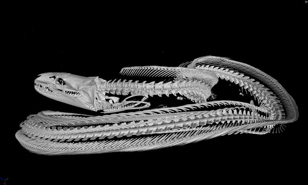
Header image: A cleared and stained backbone from a hatchling skate. Red staining indicates mineralised cartilage and blue staining indicates unmineralised cartilage.
