

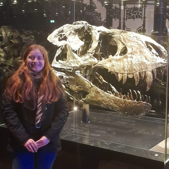
Annabel Hunt, Department of Earth Sciences, writes:
I study dinosaur skull anatomy for my PhD research at the University of Cambridge. I am a member of the Field Palaeobiology Research Group and I am supervised by Professor Daniel Field and co-supervised by Professor Steve Brusatte.
Prior to starting my PhD here in Cambridge, I studied a four-year Integrated Master’s degree in Earth Sciences at the University of Oxford. My 4th year project (supervised by Professor Roger Benson) involved using computed tomography (CT) scanning to allow me to describe various skull bones from a 254-million-year-old lizard-like reptile called Youngina capensis*. The CT scanning process produced a large number of images, and it was my job to go through each of these images (about 3000 in total!) and to manually outline the boundaries of each of the relevant skull bones. The result of this process was the generation of three-dimensional digital models of each of the relevant bones, which I could then digitally ‘zoom in’ to the regions I was interested in. Using CT scanning enabled me to identify anatomical features of Youngina that had not been previously observed, and I found this really exciting!
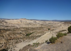
CT scanning is a fantastic tool for examining fossil material, but nothing beats the feeling of being able to physically hold fossil material in your own hands! An incredible moment for me was when I was handed a freshly excavated tyrannosaur tooth during a palaeontological excavation in the remote Badlands of Utah, and the cutting edges on the tooth felt just as sharp as they would have been when the tyrannosaur was alive over 70 million years ago!

My dinosaur research here at the University of Cambridge mostly involves using CT scans to allow me to reveal novel features of dinosaur skulls. I examine both extinct (such as T. rex) and living dinosaurs (yes, birds are dinosaurs!) to enable me to piece together the evolution of various aspects of the dinosaurian skull from extinct to living forms.
* This is the publication that resulted from my research: Hunt, A. K., Ford, D. P., Fernandez, V., Choiniere, J. N., & Benson, R. B. J. (2023). A description of the palate and mandible of Youngina capensis (Sauropsida, Diapsida) based on synchrotron tomography, and the phylogenetic implications. Papers in Palaeontology, 9(5). https://doi.org/10.1002/spp2.1521

One thought on “Imaging Dinosaurs”