

Dr. Elizabeth Marin, Department of Zoology, writes:

I have always been fascinated by insects and raised silkworms and praying mantids as a child. At university, I learned to use the fly Drosophila melanogaster to identify genes used to build animal bodies, including those of humans. Scientists have been working with this insect for over a century, describing gene mutations that change its appearance or behaviour and even developing methods to turn genes of interest on and off in specific types of cells to test their functions.
For my PhD thesis, I studied the neurons that the fly uses to discriminate between odours and the genes that remodel these neurons during metamorphosis, when a larva transforms into an adult. As a postdoctoral researcher I began working on the ventral nerve cord, the insect equivalent of our spinal cord, which is essential for most behaviours including walking, flight, and copulation. Specifically, I studied the Hox genes, a highly conserved set of genes that change the development of cells according to their locations in the body. Mutations in these genes can make flies grow legs from their heads or cause limb malformations in humans, and they also tell specific neurons whether to live or die, according to where they will be needed.
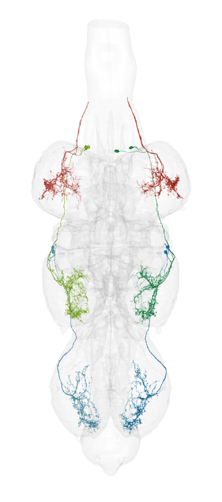
I joined Greg Jefferis’ Drosophila Connectomics group in Zoology in 2017 to help create fly brain and nerve cord connectomes – detailed atlases of every neuron and how they connect to one another – based on electron microscopy. My first study focussed on the neurons that help insects sense changes in temperature and humidity. I followed this by studying the brain structures used to associate what insects see or smell with experiences of pain or pleasure. Later I led a project on the ventral nerve cord, which was very exciting for me because I could finally see the adult forms of the cells that I had studied as a postdoc. Many of these individual cells could be found in all three of the nerve cord segments that control the fly’s pairs of legs, and we could see that their patterns of connectivity to other neurons were the same as well.
Thanks to technical advances in the field that reduce the difficulty and cost of data collection, we are now working on whole Drosophila melanogaster nervous systems as well as another fly species and an ant. I recently applied with three colleagues for a large grant to create and analyse a brain connectome for Aedes aegypti, the yellow fever mosquito. My dream is to understand, at the cellular and molecular level, how insect nervous systems have diversified through evolution to serve their endless forms and behaviours.

One thought on “Constructing Connectomes”